Optoretinography (ORG): Noninvasive Interrogation of Retinal Cell Dynamics in Response to Visual Stimulation
We employed phase-sensitive optical coherence tomography to detect nanoscopic retinal deformations in living retinas (Invited review in BOE, 2025 · My PhD thesis). Using a customized subpixel image-registration method (J. Phys. D: Appl. Phys., 2025) and an unsupervised learning approach, we successfully demonstrated multilayered ORG responses in living rat eyes over tens of seconds. These responses involved the inner and outer segments of rod photoreceptors, the retinal pigment epithelium, and the subretinal space (Nat. Commun., 2024). We also demonstrated rapid (<5 ms), nanometer-scale mechanical deformations in both human and rat rod photoreceptors upon rhodopsin activation, the earliest stage of vision (Light Sci. Appl., 2026). The rapid contraction of the rod outer segment was explained as an electromechanical manifestation of the rod early receptor potential generated in disk membranes (Nat. Commun., 2025· Review on electromechanical deformation of biological neurons, 2025). More recently, we measured multilayered ORG responses in healthy human participants and patients with retinitis pigmentosa. We further explored the use of these multilayered ORG responses, driven by intrinsic light-evoked forces in cone outer segments, to investigate retinal biomechanical properties (BioRxiv, 2026).
I believe that ORG not only helps advance our understanding of retinal physiology and biophysics, but also holds great potential for the early diagnosis of retinal degenerative diseases and for monitoring the efficacy of emerging treatments for these conditions.
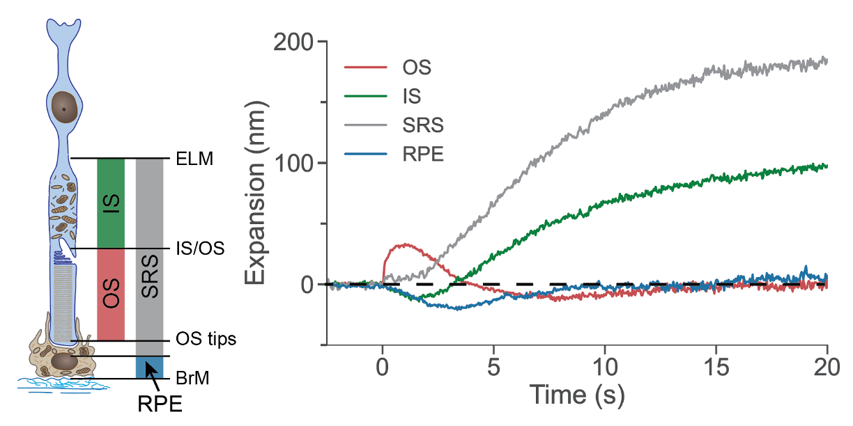
Prolonged, multilayered ORG responses in the rat retina. Adapted from Nat. Commun., 2024 & Biomed. Opt. Express, 2025.
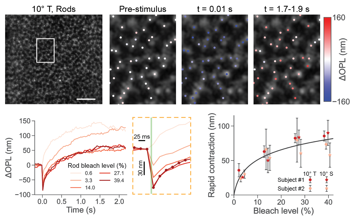
Rapid contraction in the human rod outer segment in response to strong bleaching stimuli. Adapted from Light Sci. Appl., 2026.
High-Performance Optical Coherence Tomography Angiography (OCTA) and Its Applications in Retinal, Dermatologic, Endoscopic, and Brain Research
During my undergraduate studies, I developed several high-performance OCTA algorithms that distinguish dynamic blood-flow signals from the static tissue background using classifiers constructed in multidimensional feature spaces (A review on OCTA, 2021 · Opt. Lett., 2021 · Quant. Imaging Med. Surg., 2022). We also explored OCTA applications for the noninvasive assessment of blood glucose levels in the mouse retina (Biomed. Opt. Express, 2021), evaluation of port-wine stains (Quant. Imaging Med. Surg., 2021), buccal mucosa imaging (Quant. Imaging Med. Surg., 2022), and monitoring of stimulus-evoked hemodynamic responses in the brain (IEEE Trans. Med. Imaging., 2020).
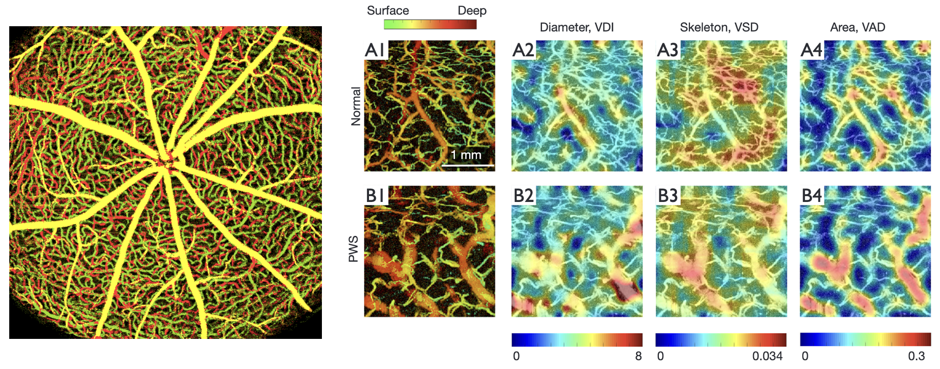
Left: Representative depth-encoded OCTA image of the mouse retina. Related methods and analyses were reported in Opt. Lett., 2021. Right: Quantitative analysis of skin vascular morphology in healthy participants and patients with port-wine stains. Reproduced from Quant. Imaging Med. Surg., 2021.